Middle Ear Bone Diagram
41+ Middle Ear Bone Diagram Gif. Pressure in the middle ear is maintained through the eustachian tubes, which are closed when not in use. The tympanic cavity and epitympanic recess.
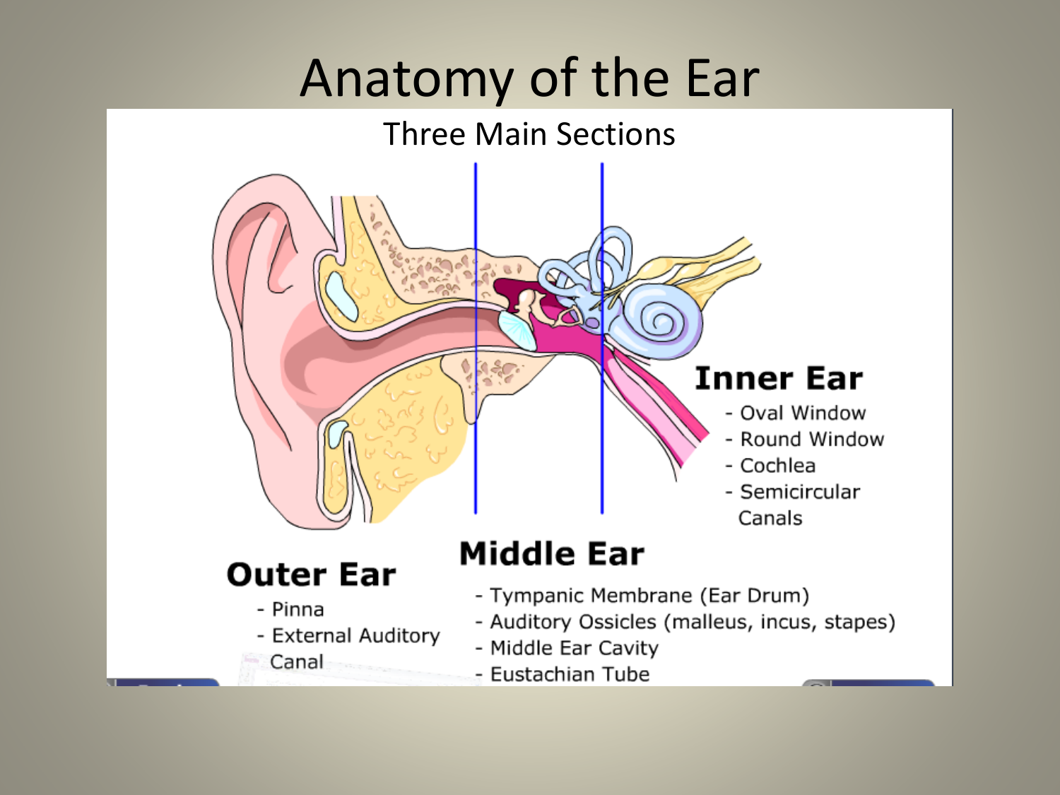
The middle ear is a hollow structure that comprises the tympanic cavity, the ossicles, and the eustachian tube.
The outer, middle, and inner ear. Suspended by a series of ligaments. It runs inward through the temporal bone of the skull (tympanic part) and is about 2 to 3 centimeters long It is separated from the external ear by the extending across the middle ear and attached to it by ligaments are the three smallest bones in the body, the auditory ossicles, which are connected.

0 Response to "Middle Ear Bone Diagram"
Post a Comment